


Daily Routines After Breast Reduction Surgery
The recovery process after breast reduction (mammoplasty) is focused on protecting the suture line, managing pain, and reducing the risk of infection. For this reason, planning your daily routines correctly is just as important as the surgical technique itself.


Rhinoplasty Prices
Who is a candidate for rhinoplasty? How is rhinoplasty performed? How are rhinoplasty costs determined? rhinoplasty Rhinoplasty is a surgical procedure performed on individuals with breathing problems and/or nasal deformities. Before rhinoplasty, the individual must not have any contraindications. It is important for the patient to prepare themselves for the change in nasal shape, have realistic expectations about the surgery, and be aware of their unique anatomical character


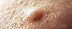
Treatment of epidermoid cyst
What is an epidermoid cyst? What are the symptoms of an epidermoid cyst? How is an epidermoid cyst treated? What is an epidermoid cyst? An epidermoid cyst is a slowly growing, usually well-circumscribed, round-shaped lesion under the skin. Unless infected, it is painless and does not negatively affect general health. If signs of infection develop, it can cause pain and discomfort, or cause cosmetic problems. Where does an epidermoid cyst occur? It occurs when the keratin in o


How long after breast augmentation can I swim?
One of the most frequently asked questions after breast augmentation surgery is when it's safe to swim in the sea or pool. This question...