
Skin Tumors
(Benign Lesions and Skin Cancers)
All lesions that originate from the skin and its appendages are called skin tumors. They can be present from birth or appear later, anywhere on the body; they may be skin-colored or a different color, flat or raised, or located under the skin. Most skin tumors are benign. However, examining every new or changing lesion allows skin cancers to be caught at an early stage.
In short: Skin tumors are divided into three groups: benign lesions (moles, cysts, lipomas, seborrheic keratoses), precancerous lesions and skin cancers (basal cell carcinoma, squamous cell carcinoma, melanoma). Treatment usually involves surgical removal and pathological examination. Early diagnosis determines both the success of treatment and the cosmetic result.
Signs of Skin Tumors
-
New spot, bump or lump: newly appeared or noticed
-
Growth: gradually increasing in size
-
Change: shape, color, texture or border changing over time
-
Irregular edges: blurred, notched or asymmetric appearance
-
Color variation: more than one shade in the same area (brown, black, red, blue-gray, whitish)
-
Persistent crusting/bleeding: bleeding on its own or bleeding easily
-
Wound-like appearance: a sore that does not heal for more than 3–4 weeks
-
Oozing/discharge: local discharge, moisture, worsening crust
-
Itching, burning or pain: especially new complaints that do not go away
-
Thickening/hardening: noticeable firmness or elevation when touched
-
Wart-like or rough appearance: an increasingly rough surface
-
Raised “tumor-like” lump: a nodule or lesion that becomes noticeable quickly
-
Swollen lymph nodes (in advanced cases): persistent swelling in the armpit, neck or groin
Classification of Skin Tumors
Skin tumors can generally be classified as benign or malignant. Benign skin tumors usually grow locally, do not spread significantly to the surrounding tissues (do not metastasize), and in most cases can be completely cured when removed surgically.
Malignant skin tumors, on the other hand, behave aggressively, can infiltrate the surrounding tissue and, in some types, can spread (metastasize) to the lymph nodes or distant organs.
Benign Skin Tumors
Benign skin tumors can originate from different cells of the skin or from the skin appendages.
Epidermal lesions (originating from keratinocytes)
-
Seborrheic keratosis (senile wart)
-
Viral warts (verruca)
-
Epidermal cysts
-
Precancerous epidermal lesions
Melanocytic lesions
-
Nevus (melanocytic nevi, moles)
-
Blue nevus
→ Details: Blue nevus (in Turkish) -
Dysplastic (atypical) nevus
-
Spitz nevus
-
Congenital melanocytic nevus
-
Nevus of Ota
-
Conjunctival and mucosal nevus
Dermal/Mesenchymal Lesions
-
Fibroma (dermal fibroma)
-
Lipoma (fatty tissue)
-
Hemangioma / vascular malformations (benign vascular lesions)
-
Lymphangioma
-
Leiomyoma (smooth muscle origin)
Neural lesions
-
Neurofibroma
-
Traumatic neuroma
-
Schwannoma
Skin appendage (hair follicle and sweat gland) lesions
-
Trichoepithelioma (hair follicle)
-
Pilomatricoma
-
Syringoma (eccrine sweat gland)
-
Eccrine poroma
-
Cylindroma
-
Apocrine cystadenoma
Hamartomatous lesions and other lesions
-
Xanthoma (yellowish lesions caused by fat deposits)
-
Hamartomas (e.g. lesions associated with certain syndromes)
-
Xanthelasma
-
Glomus tumor
-
Granular cell tumor

Melanocytic nevus

Seborrheic keratosis
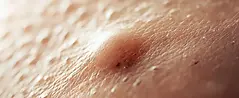
Epidermoid cyst / Epidermal cyst

Lipoma

Congenital melanocytic giant nevus
Treatment of Benign Skin Tumors
The treatment is decided according to the size, type and location of the lesion, taking into account any functional or cosmetic concerns.
Non-surgical methods
-
Observation (follow-up): Lesions that cause no symptoms and do not grow can be checked regularly.
-
Curettage (scraping) / electrocautery (burning): Can be used for superficial or easily accessible lesions.
-
Cryotherapy (freezing): Used for some benign lesions (e.g. some wart-like conditions).
-
Laser treatments: May be preferred especially for vascular lesions or suitable lesion types.
-
Topical medications (for some types): For some superficial lesions, treatments such as creams or solutions can be used if the doctor considers them appropriate.
Surgical treatment
We perform surgical treatment for benign skin tumors and lesions to remove the lesion completely, to make a definitive diagnosis (by sending it to pathology), or to relieve symptoms or a cosmetic concern. The methods we use most often:
-
Shave excision (superficial shaving): In some superficial, well-defined lesions, we can remove the lesion by shaving it off at a level above the skin. In suitable cases, it is quick and may leave less of a scar.
-
Punch biopsy / excisional biopsy (removal for diagnosis): If a lesion looks “benign” but we cannot be sure, we may need to take a sample for diagnosis or, especially for small lesions, remove almost the entire lesion and send it to pathology.
-
Excision (complete removal): Benign lesions are removed with a 1–2 mm margin of healthy tissue; if cancer is suspected, this safety margin is widened. We always send the removed tissue to pathology.
-
Reconstruction (wound closure): After removal, the wound can be closed with stitches; for large wounds or functionally important areas, we use plastic surgery repair methods such as flaps or skin grafts.
Skin Cancers (Malignant Tumors)
The most common skin cancers are basal cell carcinoma (BCC) and squamous cell carcinoma (SCC), which originate from keratinocytes, and malignant melanoma, which originates from melanocytes. Rarer types include Merkel cell carcinoma, skin lymphomas (primary cutaneous lymphomas), sarcomas (e.g. Kaposi sarcoma) and dermatofibrosarcoma protuberans.
Basal Cell Carcinoma (BCC)
-
Originates from keratinocytes in the lower layer of the epidermis
-
Usually appears after the age of 40
-
More common in men and in people with fair skin
-
Appears especially on the sun-exposed face, most often on the nose (in Turkish)
-
Can appear as a raised, crusted sore or an ulcerated, bleeding lump. Over the years, it slowly grows and begins to affect the surrounding tissues.
-
Grows slowly and has a low potential to spread
-
Treatment:
-
It can be completely cured by surgical removal.
-
To prevent recurrence, the tumor is removed together with the surrounding healthy tissue. This safety margin is usually 4 mm for low-risk tumors and wider for large or high-risk tumors.
-
Tumors removed from the face can be repaired with flaps prepared from the surrounding tissues or with grafts, known as skin grafts.
-
Basal cell carcinoma very rarely spreads to distant organs; however, if left untreated, it grows and affects the surrounding tissues.
Squamous Cell Carcinoma (SCC)
-
Can occur anywhere on the body
-
More common in sun-exposed areas such as the face, lower lip (in Turkish), ears and back of the hands
-
Smoking and excessive sun exposure trigger its development.
-
Can develop on actinic keratosis, old burn scars and long-standing non-healing wounds.
-
Squamous cell carcinoma grows slowly but has the potential to spread to the surrounding tissues.
-
It can spread especially to the regional lymph nodes and, in advanced stages, to distant organs such as the lungs.
-
Treatment:
-
The tumor is surgically removed together with the surrounding healthy tissue.
-
If needed, the tissue loss is repaired with a flap or skin graft.
-
If it has spread to the lymph nodes in the neck, a neck dissection or radiotherapy to the neck may be needed.
-
Squamous cell carcinoma can be more aggressive than BCC; follow-up after treatment is very important to detect recurrence early.

Malignant Melanoma
-
Malignant melanoma is the most feared type of skin cancer.
-
It is less common than BCC and SCC, but more aggressive.
-
When caught at an early stage, it can be treated with surgery; when advanced, it can spread to the lymph nodes and internal organs and become life-threatening.
-
It appears as a new dark lesion or one that changes in shape or color; it can develop from an existing mole or from normal skin.
-
In men, it most often appears on the back; in women, on the legs. It can occur anywhere on the body, including under the nails, on the palms and soles, and on the mucosa of the mouth, nose and genital area.
-
Treatment:
-
The tumor must be removed with a 0.5–2 cm margin of healthy tissue, and the resulting tissue loss repaired with plastic surgery methods.
-
When needed, the lymph nodes are checked with a sentinel lymph node biopsy; if they are involved, these lymph nodes are also removed.
-
Afterwards, additional cancer treatments may be needed depending on organ involvement and lymph node spread.
-
Although malignant melanoma is classified in different ways, the most important factor that determines the success of treatment is the thickness of the tumor (Breslow depth).
Precancerous Lesions
When these lesions are treated early, they can be controlled before progressing to cancer. If left untreated, they can turn into skin cancer over time.
-
Actinic keratosis: Rough, scaly, pink-brown patches on skin that has been exposed to the sun for a long time. It is the most common precursor of squamous cell carcinoma. People with actinic keratosis also have a higher risk of other skin cancers, including basal cell carcinoma, because of sun damage; regular skin checks are therefore recommended.
-
Bowen’s disease: The earliest stage of squamous cell carcinoma, limited to the top layer of the skin. If left untreated, it can progress to deeper tissues.
-
Keratoacanthoma: A raised lesion with a crusted center that grows rapidly over weeks. Because it is difficult to distinguish from squamous cell carcinoma, it is surgically removed and sent to pathology.
Frequently Asked Questions
Will there be a scar after a skin tumor is removed?
In most cases, a stitching/reconstruction plan is made according to the size and location of the lesion and the extent of removal; the aim is the best functional and cosmetic result. The scar depends on the extent of the surgery.
How can you tell whether the removed tissue is cancer?
The removed tissue is sent to pathology, examined under a microscope with special staining methods, and the diagnosis is made based on this examination.
What wound closure methods can be used?
Direct stitching for small lesions; for larger areas, skin grafts, local flaps or distant tissue transfers can be used.
What follow-up and care are needed after surgery?
After surgery, dressing and stitch checks, sun protection, wound follow-up for possible infection or blood supply problems, and check-ups planned according to the pathology result are carried out.
Is the belief that “cutting a skin lesion turns it into cancer” true?
Absolutely not. Removing skin lesions generally does not cause cancer. On the contrary, surgically removing a suspicious lesion often allows a diagnosis to be made or, if it is malignant, early diagnosis and treatment.
Can skin cancer be completely cured?
Basal cell carcinoma, as well as squamous cell carcinoma and melanoma caught at an early stage, are mostly cured completely when surgically removed with appropriate margins. In advanced stages, additional treatments may be needed alongside surgery; that is why early diagnosis is very important.
Information and Booking
When skin tumors are noticed early, the chance of successful treatment is much higher. If you have a new, growing or bleeding lesion, or one that changes in shape or color, I recommend that you be examined without delay. You can send a photo of the lesion to our WhatsApp line for a preliminary assessment. If you live abroad, this also helps us plan your trip and your stay in Türkiye.
This page was prepared and reviewed by Op. Dr. Nurgül Altuntaş .
Last update: November 2026